
Lipofibromatosis : Microscopic Features

slide 42 of 60


Comments:
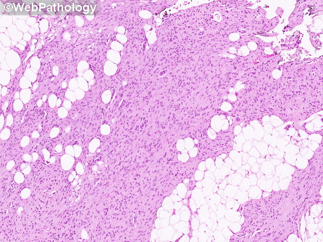
Microscopic Features: More established lesions of lipofibromatosis consist of short, intersecting fascicles of mature-appearing spindle-shaped fibroblasts that lack cytologic atypia or increased mitotic activity. The surrounding skeletal muscles, adipose tissue and fascial planes are often infiltrated. Mature adipose tissue is an integral part of the lesion. Univacuolated cells resembling lipoblasts may be seen at the interface between mature fat and fibroblastic areas (see next image). Melanin pigment may be present within the cells.
slide 42 of 60