
Ischemic Fasciitis : Microscopic

slide 8 of 12


Comments:
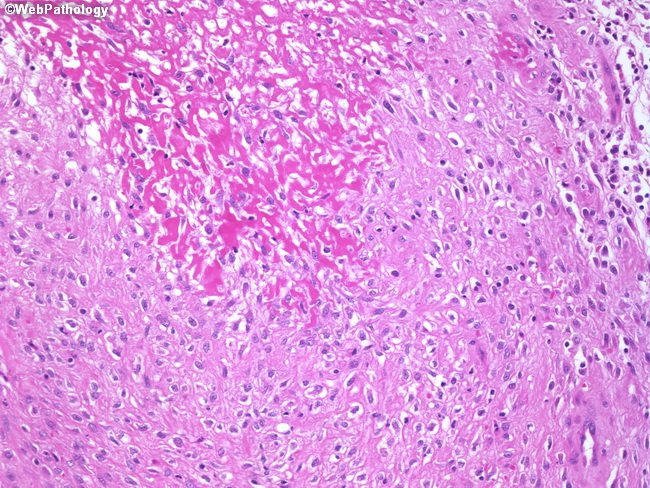
Ischemic fasciitis shows a zonal pattern which is obvious at low magnification. There is a central area of fibrinoid necrosis surrounded by a zone of vascular proliferation and ganglion-like myofibroblasts. The capillary caliber blood vessels may be lined by plump atypical endothelial cells. The blood vessels at the periphery of the lesion may show fibrin thrombi, acute inflammation, and perivascular hyalinization. Mitotic activity is increased, but atypical mitoses are not seen. Additional findings include foreign body giant cells and extravasated red blood cells.
slide 8 of 12