
Hibernoma : Microscopic Features

slide 10 of 27


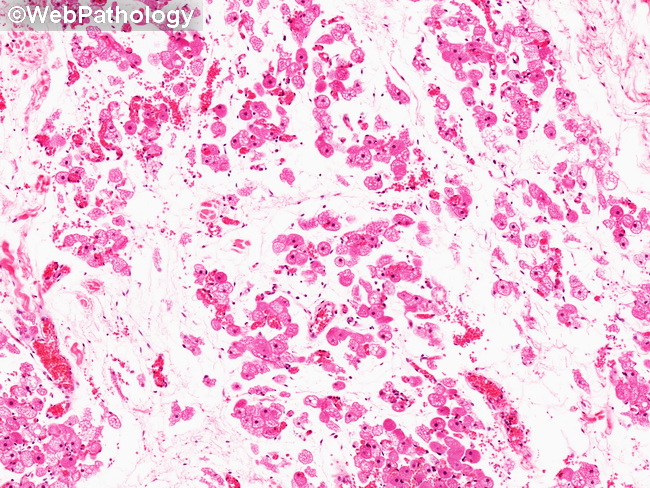
Comments:
Microscopic Features: About 9-10% of hibernomas show prominent myxoid change. The tumor cells are present singly or in small clusters in a myxoid stroma. Rare hibernomas (2%) have a spindle cell morphology. They tend to occur on scalp and back. Such cases have to be differentiated from spindle cell lipomas.
slide 10 of 27