
Spindle Cell Lipoma : Microscopic

slide 9 of 36


Comments:
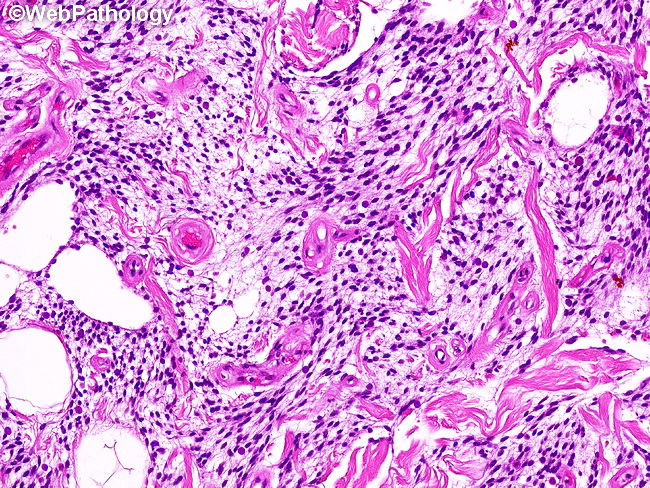
Microscopic Features: The spindle cells are arranged in short, irregular fascicles in a fibromyxoid matrix often containing thick collagen bundles. There may be prominent nuclear palisading, similar to that seen in schwannomas. Some tumors are sparsely cellular and contain abundant myxoid matrix, mimicking a myxoma. The background matrix contains numerous mast cells. Foci of osseous or cartilaginous metaplasia may be present.
slide 9 of 36