
Nodular Fasciitis : Immunohistochemistry


Comments:
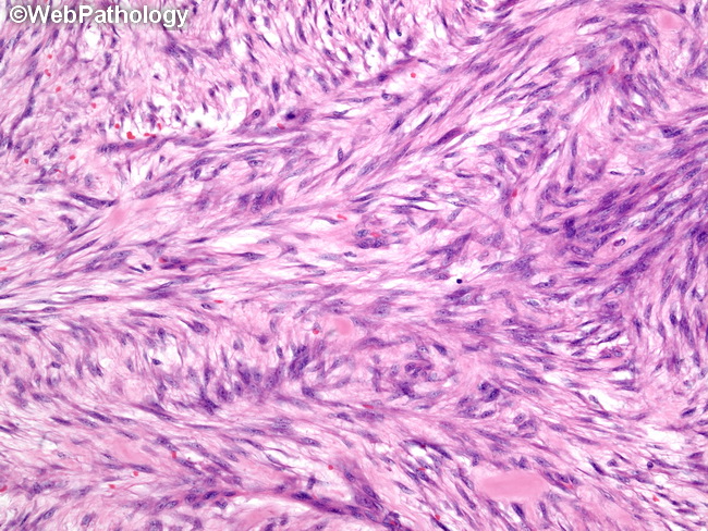
The lesional cells of nodular fasciitis are uniform, plump, immature-appearing fibroblasts and myofibroblasts resembling those seen in tissue culture. They are arranged in short, intersecting fascicles with a storiform growth pattern. The cells have oval, uniform nuclei with prominent nucleoli. Mitotic activity is brisk, but there are no atypical forms. Scattered lymphocytes, extravasated red blood cells (and microhemorrhages), foamy histiocytes, and multinucleated giant cells are also present. Immunohistochemistry: The myofibroblasts show immunoreactivity for smooth muscle actin and muscle-specific actin. There is no positivity for desmin or caldesmon. The negativity for beta-catenin is a useful feature distinguishing nodular fasciitis from fibromatoses (which are consistently positive for beta-catenin in the nucleus). Cytokeratin and S-100 are also negative.