
Nodular Fasciitis : Differential Diagnosis


Comments:
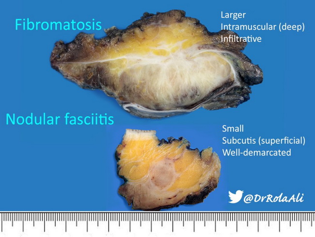
Nodular fasciitis resembles numerous benign mesenchymal lesions. Taken out of context, it is frequently misdiagnosed as a sarcoma, given its hypercellularity, rapid growth, irregular infiltrating borders (not all cases), and increased mitotic activity. Following entities enter into the differential diagnosis: Myxoma: Both nodular fasciitis and myxoma have abundant myxoid matrix. Myxoma is comparatively paucicellular and poorly vascularized. It also lacks the organization as well as heterogeneity of nodular fasciitis. Fibrous histiocytoma: Fibrous histiocytoma is usually centered in dermis (an uncommon location for nodular fasciitis), shows epidermal hyperplasia, and is poorly-circumscribed. The cell population is polymorphic and consists of spindle and round cells, chronic inflammatory cells, foamy histiocytes, siderophages, and Touton giant cells. Storiform growth pattern is more pronounced than that in nodular fasciitis. Fibrous histiocytoma stains strongly for factor XIIIa, where as nodular fasciitis shows diffuse positivity for SMA. Fibromatosis: Fibromatosis is a much larger, poorly-circumscribed, and an infiltrative lesion as compared to nodular fasciitis (see gross images). The lesional fibroblasts are arranged in long intersecting fascicles. Mitotic activity is not as high as in nodular fasciitis. Strong nuclear expression of beta-catenin is a diagnostic feature of fibromatosis and it is absent in nodular fasciitis. Other benign conditions that should be considered include neurofibroma and spindle cell lipoma. Fibrosarcoma: Fibrosarcomas are large, deeply-located masses of longer duration. They are densely cellular and consist of intersecting bundles of spindle shaped cells in a herring bone pattern. There is greater degree of cellular pleomorphism, nuclear hyperchromasia, and brisk mitotic activity (including atypical forms). Myxofibrosarcoma: The patients are usually older than 50 years and the lesion is greater than 3 cm in size. Tumor cells show greater nuclear pleomorphism, arborizing vasculature, and atypical mitotic figures. Foci of high-grade pleomorphic sarcoma are frequently present.
Image courtesy of: Rola Ali, MD, FRCPC, Associate Professor of Pathology, Kuwait University, Kuwait; used with permission.