
Nodular Fasciitis : Related Lesions


Comments:
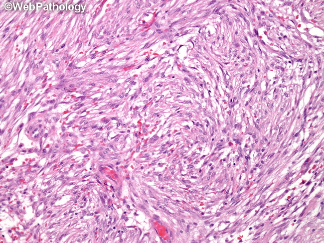
Ossifying fasciitis, panniculitis ossificans, and fibroosseous pseudotumor of digits are lesions that share features with both nodular fasciitis and myositis ossificans. Ossifying fasciitis has nodular fasciitis-like features along with metaplastic bone. Unlike myositis ossificans, it does not show zonal maturation. When arising from periosteum, it is referred to as parosteal fasciitis. Intravascular fasciitis: It is a rare, nodular fasciitis-like lesion involving small or medium-sized vessels (veins > arteries). It usually presents as a solitary, small, slow-growing, painless, subcutaneous mass in young patients. The most common location is upper extremity. Other sites include head and neck, trunk, lower extremities, and oral cavity. Cranial fasciitis is a rapidly growing myofibroblastic proliferation involving soft tissues of the scalp and the underlying cranium. It is a benign reactive process arising from the galea aponeurotica (epicranial aponeurosis). It is seen mainly in infants under 1 year of age. Microscopically, it resembles nodular fasciitis and is composed of fibroblasts and myofibroblasts in myxoid or hyalinized matrix. Osseous metaplasia may be present. This image of nodular fasciitis shows fibroblasts and myofibroblasts arranged in short fascicles with a storiform growth pattern.