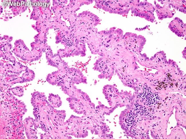
AdenoCIS of Lung : Non-Mucinous


Comments:
Pulmonary adenocarcinoma-in-situ (AIS) is usually discovered incidentally in the peripheral lung on chest CT performed for other medical reasons. It has a ground-glass or soap bubble appearance and lacks solid component on CT. The previously used term for this entity is bronchioloalveolar carcinoma which is no longer used in the WHO 2015 Classification of Lung Tumors. AIS is a slow-growing tumor. The disease-free survival is 100% if it is completely resected. AIS is not included in the current TNM classification. The image shows cuboidal type II pneumocytes lining the preexisting alveolar structures. There is widening of the alveolar septa due to sclerosis, elastosis and chronic inflammation. The pneumocytes have low-grade cytologic features, including small bland nuclei with punctate nucleoli. Mitotic activity is not increased. AIS expresses TTF-1 and Napsin A. It must be distinguished from minimally-invasive adenocarcinoma by thorough sampling and careful search for invasive foci.


