
Infected Hydrocele

slide 41 of 61


Comments:
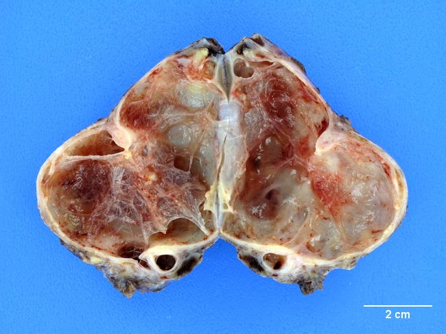
Hydroceles may be complicated by hemorrhage or infection. The image shows an infected hydrocele removed from a patient with history of epididymo-orchitis. The specimen was filled with blood-tinged, cloudy mucoid fluid. The inner wall is rough and contains numerous delicate fibrous septa dividing the cyst into locules. The wall thickness ranged from 0.1 cm to 1.0 cm secondary to chronic inflammation and fibrosis.
slide 41 of 61