
Paget Disease of Bone : Radiology


Comments:
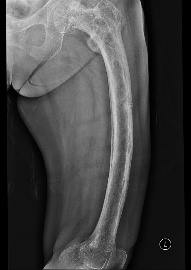
Paget Disease of Bone (PDB) - Imaging Studies: In the later stages of PDB, when osteoblastic activity takes over, there is enlargement and widening of the bone with marked cortical thickening. The cancellous bony trabeculae become thicker, coarser, and irregular. There is loss of corticomedullary demarcation. The periosteal and endosteal surfaces also become rough and irregular. Case History: This plain X-ray (frontal view) is from an 80 y/o female who presented with chronic severe left hip pain, progressive difficulty walking and deformity of the upper left thigh. There was no history of trauma. Initial imaging studies showed diffuse cortical and coarse trabecular thickening with bowing of left femur. There were multifocal osteolytic defects along the cortex. In addition, there was a transverse insufficiency fracture (banana fracture) at the lateral aspect of the cortex of the femur (see annotated next image). Case courtesy of Dr Salman S. Albakheet, Radiopaedia.org. From the case rID: 80359