
Paget Disease of Bone : Diagnosis


Comments:
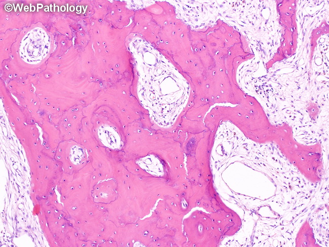
Paget Disease of Bone (PDB) - Diagnosis: PDB is often clinically silent and discovered incidentally on imaging studies or an investigation triggered by an isolated finding of elevated serum alkaline phosphatase (SAP) level. Other conditions that may raise SAP include vitamin D deficiency, hyperparathyroidism, hyperthyroidism, renal osteodystrophy, and malignancy. Plain X-rays of the abdomen, femurs, tibias, skull and facial bones are recommended as the initial diagnostic screening test in patients suspected of having PDB on biochemical or clinical grounds. Xrays of the affected bone/s show the characteristic features as discussed previously. Isotope bone scans are quite sensitive and accurate diagnostic tool and show increased uptake in active stages of the disease. Bone scans may be negative in sclerotic (burnt-out) phase of the disease. CT scans and MRI are not recommended for the initial diagnosis but are useful for the assessment of complications of PDB such as basilar invagination, spinal stenosis, and osteosarcoma. Bone biopsy may be necessary in some cases to confirm the diagnosis. This image shows some of the classic features of late stage PDB - irregular, thick, plate-like trabeculae; prominent basophilic cement lines; and fibrovascular tissue replacing bone marrow.