
Paget Disease of Bone : Microscopic


Comments:
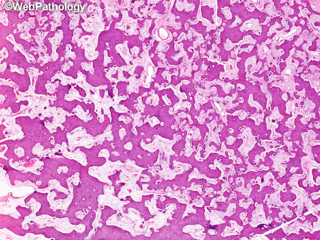
Paget Disease of Bone (PDB) - Microscopic Features: The microscopic appearance of PDB depends upon the stage of the disease. Three stages have been described - osteocytic (early, acute phase), mixed osteolytic and osteoblastic (intermediate phase) and sclerotic phase (late, burnt-out). Osteolytic Phase: There is excessive bone resorption by osteoclasts that are larger than normal and have more numerous nuclei. The frenetic osteoclastic activity creates irregular, variably sized resorption cavities with scalloped edges in the cortical bone whereas cancellous bone shows irregular, anastomosing and branching trabeculae as seen here. The bone marrow shows peritrabecular fibrosis. There are no inflammatory cells. It is not uncommon to see all phases of disease simultaneously in a single specimen from established cases of PDB. The picture in acute osteolytic phase of PDB resembles hyperparathyroidism (hyperPTH). However, the osteoclasts in PDB are larger than those seen in hyperPTH and have upwards of 10-20 nuclei. HyperPTH shows tunnelling resorption, a feature absent in PDB.