
Myositis Ossificans - Differential


Comments:
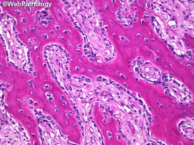
Differential Diagnosis (continued): Myositis ossificans (MO) has overlapping features with osteosarcoma. Biopsies taken from the cellular center during the early phase of MO are especially challenging to differentiate from extraskeletal osteosarcomas. Careful evaluation of clinical and radiologic findings is essential. Broadly speaking, MO is most mature at the periphery and primitive-appearing in the center. Osteosarcomas show the reverse pattern - more mature in the center and primitive at the periphery. Features supporting extraskeletal osteosarcoma: older patients (6th to 7th decades; rare in young); may show reverse zoning with osteoid or bone in the center and atypical spindle cells at the periphery; irregular lace-like osteoid (rather than thicker trabecular osteoid) lined by hyperchromatic pleomorphic cells; greater degree of cellular atypia; invasion/destruction of adjacent tissues; and atypical mitoses. About this image: Mineralized osteoid trabeculae rimmed by plump osteoblasts near the periphery of a lesion of myositis ossificans.