
Myositis Ossificans - Differential


Comments:
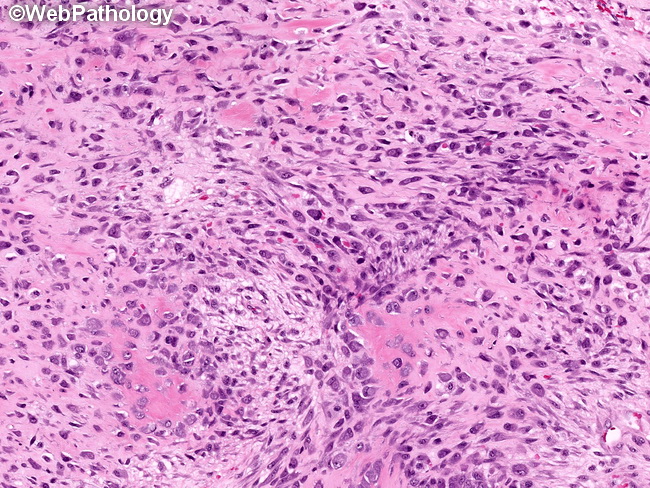
Differential Diagnosis: When there is characteristic history and the zonal pattern on morphology and imaging studies, the diagnosis of myositis ossificans (MO) is straightforward. However, depending upon the stage of the lesion at the time of biopsy or excision, it can mimic several malignant and benign processes such as: extraskeletal osteosarcoma (most important), parosteal osteosarcoma, metastasis (osteoblastic carcinoma, melanoma), and benign reactive processes (nodular fasciitis, proliferative myositis, posttraumatic periostitis, exuberant fracture callus, or even a soft tissue abscess). Proliferative myositis (PM) may show small foci of osteoid. Features favoring PM include the presence of ganglion-like plump fibroblasts. Posttraumatic periostitis is attached to the bone with a broad base. Exuberant callus is associated with a fracture. About this image: This image taken from the interface between the innermost and the intermediate zones shows transition between fibroblastic and osteoblastic areas in myositis ossificans. The plump reactive fibroblasts/myofibroblasts are undergoing metaplasia to osteoblasts and there is appearance of osteoid matrix (homogenous pink areas). Myositis Ossificans vs Osteosarcoma is discussed in the next image.