
Myositis Ossificans - Morphology


Comments:
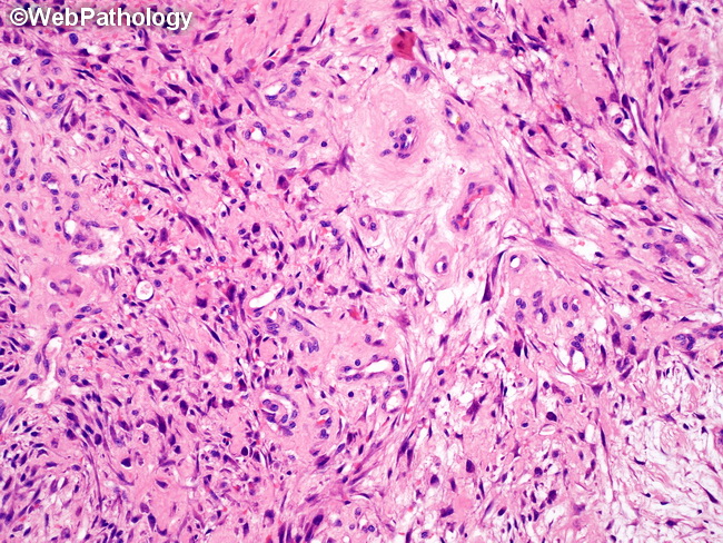
Microscopic Features: The zonal pattern is an important diagnostic clue for myositis ossificans (MO). In specimens that are at least 3-4 weeks old, the zonal pattern is well-established and consists of 3 zones reflecting different levels of cellular maturation as described in the previous two images. The appearance shown here is characteristic of early phase of MO as well as the innermost zone of the more mature lesions of MO. It resembles nodular fasciitis, granulation tissue or a sarcoma and consists of an irregular proliferation of immature fibroblastic/myofibroblastic cells in a highly vascular, collagenous stroma. The plump spindle cells show mild cytologic atypia. There are numerous mitotic figures; however, atypical forms should not be present. The myofibroblastic tissue may be admixed with chronic inflammatory cells, macrophages, multinucleated giant cells, and fibrinous material. Interstitial microhemorrhages and entrapped muscle fibers may also be present.