
Myositis Ossificans - Morphology

slide 10 of 24


Comments:
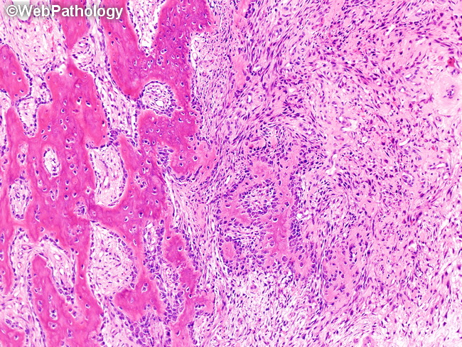
Microscopic Features: Higher magnification of the previous image showing the characteristic zonal pattern of myositis ossificans. The innermost zone (right) consists of highly cellular, immature-appearing, loosely textured, fibroblastic/myofibroblastic proliferation in a vascular, collagenous background. The intermediate zone (center of the image) shows transition from spindle cell stroma to irregularly-shaped, poorly-defined osteoid trabeculae line by plump osteoblasts. The outermost zone (left) shows osteoid trabeculae undergoing mineralization to mature into arrays of lamellar bone.
slide 10 of 24