
Aneurysmal Bone Cyst : Gross


Comments:
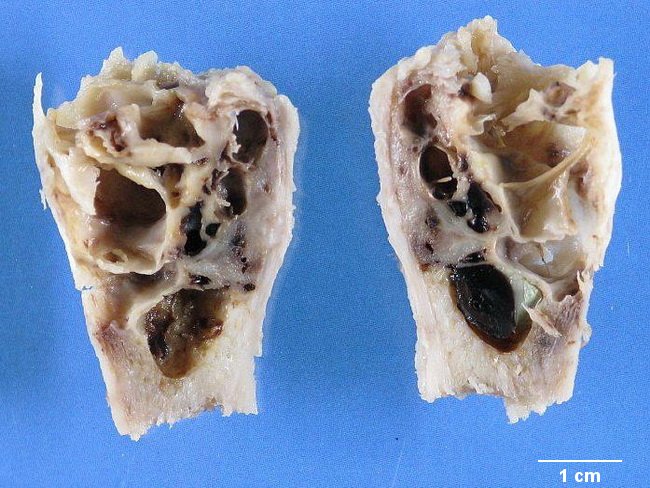
Aneurysmal Bone Cyst (ABC) - Gross Pathology: A completely resected ABC appears as a spongy, honeycombed mass of blood-filled cystic spaces separated by gray-white fibrous septa of variable thickness. The tumor causes "blow out" expansion of the involved segment of the bone with cortical thinning. The lesion may extend into soft tissues and is surrounded by a thin layer of reactive bone. Solid pink-tan areas may be present and should be thoroughly sampled to rule out any other underlying primary tumor (i.e. to differentiate between primary ABC and ABC-like changes. Rare lesions are completely solid and show no evidence of cysts or septa. They represent solid variant of ABC. Frequently, ABCs are removed by aggressive curettage and the specimen received by the laboratory consists of mushy, red-brown, hemorrhagic granular material with bone fragments. Image courtesy of Dr. Jean-Christophe Fournet, Paris, France; humpath.com; Used with permission