
Chordoma : Imaging

slide 5 of 51


Comments:
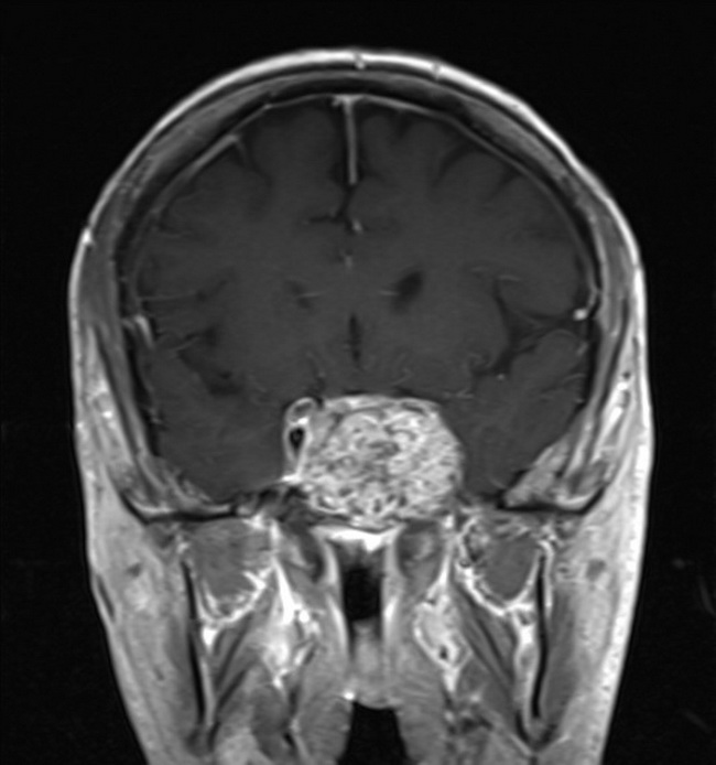
Case History: Coronal T1 MRI image showing a chordoma arising at the clivus. After sacrococcygeal region, clivus is the second most common location for chordomas. The patient was a 50 y/o male who presented with progressive headaches, diplopia and numbness on his face. Same case as the previous image. Case courtesy of Dr Dylan Kurda, Radiopaedia.org. From the case rID: 36795
slide 5 of 51