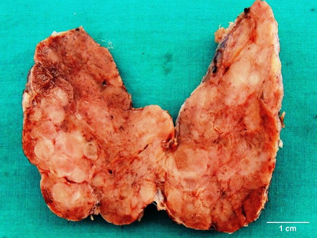
Hashimoto Thyroiditis


Comments:
This thyroidectomy specimen is from a 46 y/o female who presented with painless, gradual enlargement of thyroid gland involving both the lobes. She was hypothyroid and was positive for antithyroid antibodies. Micrscopic examination showed severe thyroid atrophy and lymphoid follicles with prominent germinal centers separated by dense hyaline fibrosis. The findings were diagnostic of Hashimoto thyroiditis. The specimen photographs shows a diffusely enlarged thyroid with ill-defined whitish nodular areas secondary to fibrosis. Image courtesy of: Dr. Sanjay D. Deshmukh, Professor of Pathology, Dr. Vithalrao Vikhe Patil Medical College & Hospital, Ahmednagar, India.


