
Hodgkin Lymphoma in Liver


Comments:
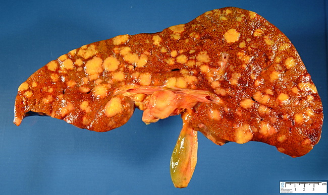
Primary hepatic lymphomas are rare. Most occur in middle-aged or elderly patients with an underlying immunodeficiency (prior organ transplantation), chronic infection (Hepatitis B, C; HIV infection; active tuberculosis), or autoimmune disorder (SLE, Sjogren syndrome; Felty syndrome). Secondary involvement of liver by lymphomas is more common. Most are diffuse large B-cell lymphomas. Other types include MALT lymphomas, Burkitt lymphoma, follicular lymphoma, peripheral T-cell lymphoma, and Hodgkin lymphoma. In 50% of cases, lymphomas form a solitary mass. In about 45% of cases, there are multiple small nodules scattered throughout the liver. In 5% of cases, there is diffuse hepatic enlargement without a dominant mass. The differential diagnosis includes hepatocellular carcinoma or metastatic carcinoma. Elevation of lactate dehydrogenase with normal CEA and AFP favors the diagnosis of lymphoma. On FNA smears, lymphomas are discohesive neoplasms. Depending upon the specific type, the lymphoid cell population may be monomorphic without significant cytologic atypia (follicular lymphomas, MALT-lymphomas, and small lymphocytic lymphomas) or composed of large atypical cells (diffuse large B-cell lymphoma - the most common lymphoma secondarily involving the liver). Lymphoglandular bodies are present in the background. FNA material is usually sufficient to perform flow cytometry or gene rearrangement studies. Cell block or needle core biopsy specimen can be used for immunohistochemical studies.The image shows Hodgkin lymphoma involving the liver. Image courtesy of Dr. Jean-Christophe Fournet, Paris, France; humpath.com; Used with permission