
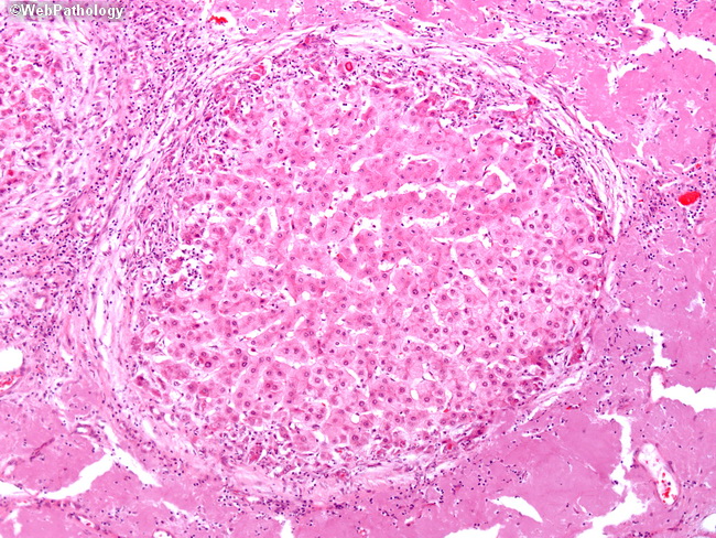
Amyloidosis of Liver

slide 11 of 15


Comments:
A higher magnification of the previous image shows marked hepatic amyloid deposition, which begins in portal vessels and hepatic sinusoids. With disease progression, the residual liver parenchyma is compressed into nodules separated by abundant amorphous, eosinophilic amyloid deposits. The presence of amyloid can be confirmed by apple green birefringence on Congo Red staining with polarized microscopy.
slide 11 of 15