
Prostatic Ductal Adenocarcinoma

slide 11 of 36


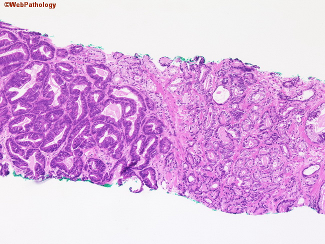
Comments:
This needle biopsy shows a focus of ductal adenocarcinoma on the left and usual acinar adenocarcinoma on the right. The two patterns were intermingled throughout the biopsies. The ductal areas are composed of larger, back-to-back glands some of which contain papillary infoldings.
slide 11 of 36