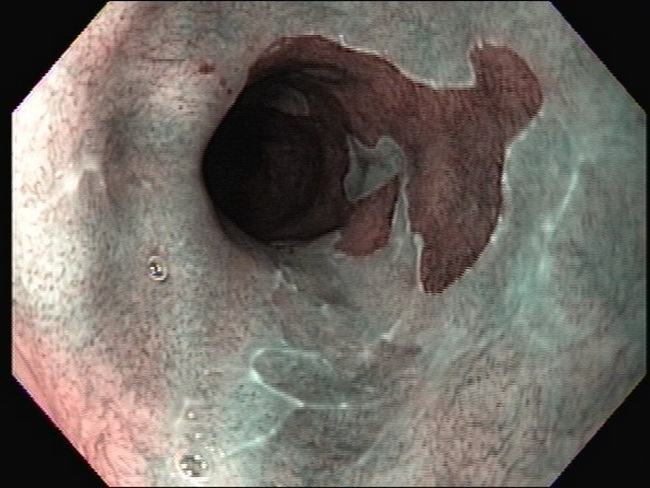
Barrett Esophagus : Clinical


Comments:
Clinical Features: The columnar epithelium of Barrett esophagus (BE) by itself causes no symptoms. Patients are usually diagnosed during endoscopic evaluation for gastroesophageal reflux disease (GERD). BE is found in almost 10% of patients with symptomatic GERD. Most patients are middle-aged or older adults (avg. age 55 years) with a chief complaint of heartburn. BE is sometimes seen in children in association with cystic fibrosis (which can cause gastroesophageal reflux) or following chemotherapy. In addition to the history of GERD, other risk factors associated with the development of BE include white race, male gender, age > 50, hiatal hernia, tobacco use, and central obesity. This endoscopic view with narrow band imaging (NBI) is from the same case as the previous image. The columnar epithelium lining the distal esophagus is clearly delineated as dark brown mucosa against normal squamous epithelium which appears greenish. Further discussion of NBI is in slide 4. Image courtesy of: Pramod Malik, MD; used with permission.


