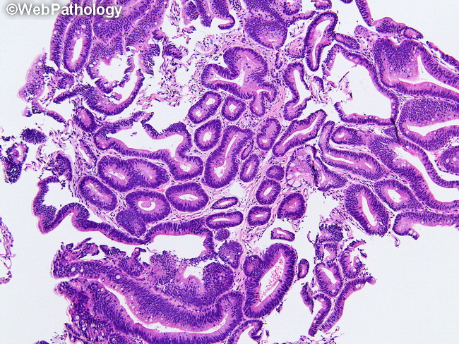
Barrett Esophagus : Low-grade Dysplasia


Comments:
Barrett Esophagus (BE) with Low-grade Dysplasia: The biopsies with low-grade dysplasia show atypical cytologic features and resemble tubular adenomas of colon. There is only minimal or no surface maturation, i.e. the cytologic atypia of the basal crypt glands is also seen in the surface epithelium. There is glandular overcrowding; however, lamina propria is still identifiable between glands. There is mild to moderate architectural distortion. The glands are lined by cells with elongated hyperchromatic nuclei, increased nuclear:cytoplasmic ratio, maintained nuclear polarity, and increased mitoses. There is nuclear membrane irregularity. Nucleoli are not prominent. There may be some nuclear stratification. In the presence of inflammation, biopsies with features of low-grade dysplasia are perhaps better classified as indefinite for dysplasia. There is considerable uncertainty about the rate of progression of LGD to adenocarcinoma, given the high degree of interobserver variation in the diagnosis of LGD. The reported rate is about 20% in 5 years. Image courtesy of: Phoenix Bell, MD.


