
Herpes Esophagitis

slide 8 of 29


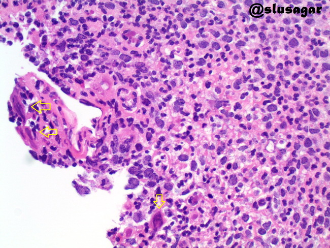
Comments:
This endoscopic esophageal biopsy is from an adult male with upper GI symptoms. Ulcers were noted at endoscopy. Biopsies showed reactive/regenerative squamous mucosa (not shown here) and ulcer. The findings were suspicious for herpes esophagitis; however, charateristic histologic findings of multinucleated giant cells with chromatin margination and Cowdry A intranuclear inclusions were not readily apparent. Yellow arrows point to possible viral inclusions. With HSV immunostain, numerous cells were positive (see next image). Case courtesy of: Ankur Sangoi, MD; used with permission.
slide 8 of 29