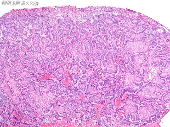
Peripheral Ameloblastoma : Microscopic


Comments:
The microscopic appearance of peripheral (extraosseous) ameloblastoma is similar to its intraosseous counterpart involving the mandible. Islands of ameloblastic epithelium in follicular or plexiform pattern are seen in the lamina propria beneath the surface epithelium. The surface mucosa is ulcerated in this image. Follicular pattern: This pattern consists of round, oval, or irregular epithelial islands that try to mimic enamel organ epithelium. The nests and islands show peripheral palisading of columnar cells with reverse polarity i.e. their nuclei are polarized away from the basement membrane. The central portion of the islands consists of angulated cells resembling stellate reticulum of the developing tooth germ. The nests are separated by mature fibrous connective tissue stroma. The plexiform pattern consists of interconnected thin lamina like strands or cords of basaloid cells, often without peripheral palisading and reverse nuclear polarity. Within these cords are more loosely arranged epithelial cells. The surrounding stroma is stroma is vascular and composed of loose connective tissue.


