
Adenoid Cystic CA : Microscopic
Home
 Head & Neck
Salivary Glands
Malignant Neoplasms of Salivary Glands - I
Adenoid Cystic CA : Microscopic
Head & Neck
Salivary Glands
Malignant Neoplasms of Salivary Glands - I
Adenoid Cystic CA : Microscopic

slide 91 of 146


Comments:
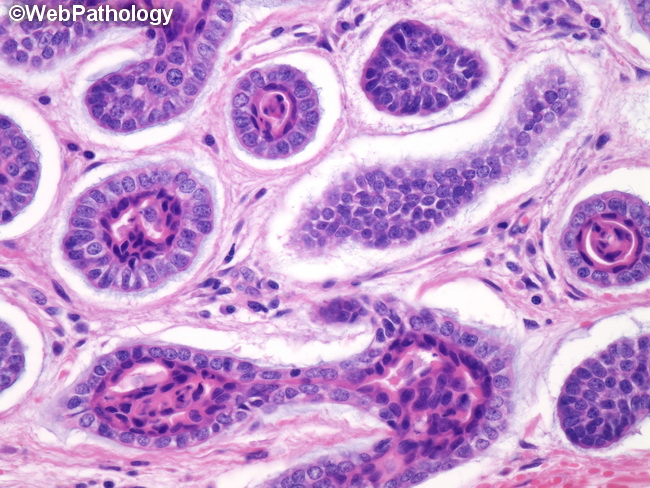
Adenoid cystic carcinoma shows an admixture of many architectural patterns, including cribriform, tubular, and solid growth with a myxohyaline stroma. There are two cell types � abundant basaloid cells with myoepithelial differentiation and eosinophilic epithelial cells with ductal differentiation. In addition to gland-like spaces (pseudocysts) filled with eosinophilic basement membrane material or basophilic myxoid material, there are also true glandular structures as shown here. They are lined by low cuboidal epithelium with eosinophilic cytoplasm and surrounded by a collar of myoepithelial cells.
slide 91 of 146