
Epithelial Cyst of Spleen


Comments:
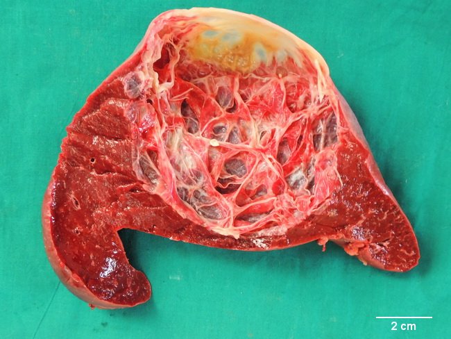
Epithelial (True) Cyst of Spleen: This splenectomy specimen is from a 40 y/o male who presented with left upper quadrant pain and low-grade fever of brief duration. There was no history of trauma. Ultrasonography showed a hyperechoic lesion suggestive of a cyst or an abscess. On CT scan, there was a well-defined hypodense lesion with no solid component or septae. The splenectomy specimen measured 16.5 x 12 x 11 cm and weighed 650 grams. The surface had a bulging yellow-white area (see previous image). The specimen contained a 9.0 cm unilocular cyst filled with about 300 ml of straw-colored fluid. The inner wall was greyish-white and heavily trabeculated as shown in this image. The cyst lining was composed of low cuboidal epithelium. Case courtesy of: Dr. Sanjay D. Deshmukh (Professor of Pathology) & Dr. J. M. Gadekar (Chief of Surgery Dept.), Dr. Vithalrao Vikhe Patil Medical College & Hospitals, Ahmednagar, India.