
M. tuberculosis lymphadenitis: Gross
Home
 Hematopathology
Lymph Node (Non-Hematopoietic)
Mycobacterial Lymphadenitides
M. tuberculosis lymphadenitis: Gross
Hematopathology
Lymph Node (Non-Hematopoietic)
Mycobacterial Lymphadenitides
M. tuberculosis lymphadenitis: Gross

slide 4 of 7


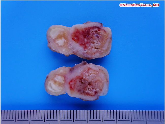
Comments:
Gross Pathology: The cut surface of the involved lymph node shows creamy-white areas of caseous necrosis with chalky white calcium deposits. With the passage of time, lymph nodes can become fibrotic, matted together, and rock-hard. On plain films, they appear as radio-opaque masses. The lymph nodes can attain large size in primary tuberculosis, especially the tracheobronchial nodes in children. The infection may involve adjacent structures resulting in strictures, abscesses, fistulas, traction diverticula and invasion of large vessels. Image courtesy of: Nejib Ben Yahia, MD, Tunisia; used with permission.
slide 4 of 7