
Mycobacterium tuberculosis lymphadenitis
Home
 Hematopathology
Lymph Node (Non-Hematopoietic)
Mycobacterial Lymphadenitides
Mycobacterium tuberculosis lymphadenitis
Hematopathology
Lymph Node (Non-Hematopoietic)
Mycobacterial Lymphadenitides
Mycobacterium tuberculosis lymphadenitis
slide 1 of 7


Comments:
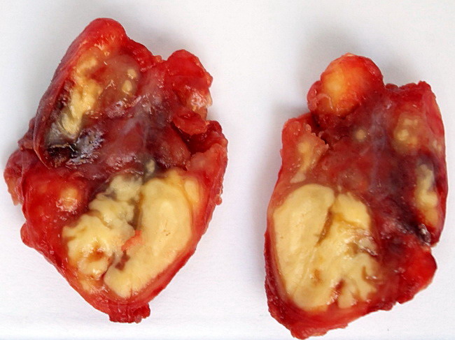
This lymph node biopsy from a young man included a couple of matted lymph nodes. The cut surface shows yellow areas of caseous necrosis. The scale is centimeters. Histology was typical but no acid fast bacilli were seen. Clinically, the matted lymph nodes may be confused with metastatic carcinoma. The most common location is cervical region. Case courtesy of Dr. Bulent Celasun, Ankara, Turkey. Used with permission.
slide 1 of 7