
Giant Cell Myocarditis

slide 8 of 16


Comments:
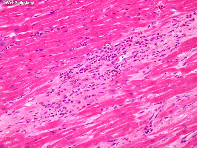
This image shows an area of eosinophilic infiltrate with focal destruction of myocardial fibers in a case of giant cell myocarditis. Multinucleated giant cells are not present in this focus. The main differential diagnostic consideration is cardiac sarcoidosis with which it may be confused; however, giant cell myocarditis does not contain well-formed granulomas of sarcoidosis.
slide 8 of 16