
Myositis Ossificans - Gross Pathology
Home
 Orthopedic
Tumor-like Lesions of Bone
Myositis ossificans
Myositis Ossificans - Gross Pathology
Orthopedic
Tumor-like Lesions of Bone
Myositis ossificans
Myositis Ossificans - Gross Pathology

slide 7 of 24


Comments:
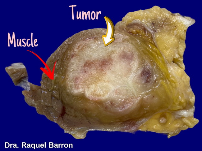
Myositis Ossificans - Gross Pathology: The excised lesions are solitary, well-circumscribed and contained within the muscle but with no connection to bone. The size ranges from 3 to 6 cm. Mature lesions have a hard, peripheral shell of ossification and a soft, gelatinous, grey-red center. They cut with a gritty sensation due to the thin shell of bone. Cystic changes may be present that can mimic aneurysmal bone cyst. The image shows a gross specimen of myositis ossificans removed from tibialis anterior muscle in a 22 y/o female. Image courtesy of: Raquel Barron, MD, Bolivia; used with permission.
slide 7 of 24