
Ependymoma

slide 6 of 48


Comments:
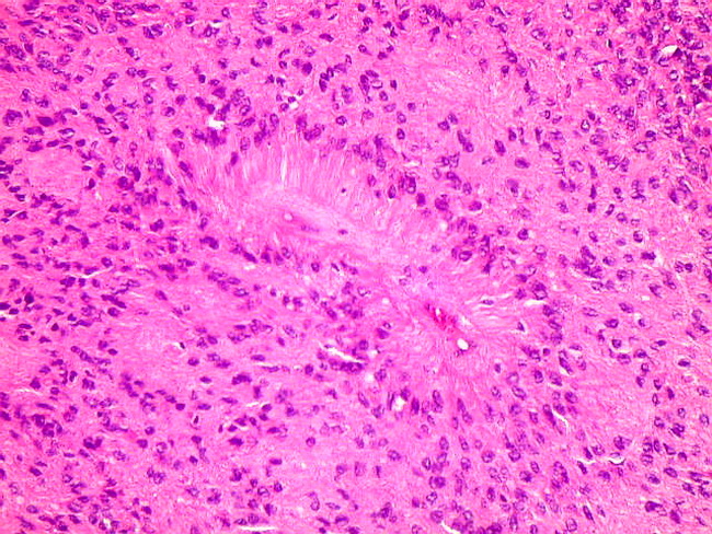
Classic ependymoma is a well-circumscribed and a cellular neoplasm with sheet-like growth pattern. The tumor cells are frequently arranged around blood vessels creating perivascular pseudorosettes as seen in the center of this image. Vast majority of ependymomas are infratentorial and involve the 4th ventricle, mostly in children. Supratentorial lesions are more likely to be cystic and show anaplasia. Courtesy of: Dr. Luciano de Souza Queiroz, Dept. of Pathology, Faculty of Medical Sciences, State University of Campinas (UNICAMP), Campinas, Sao Paulo State, BRAZIL. Additional images are here.
slide 6 of 48