
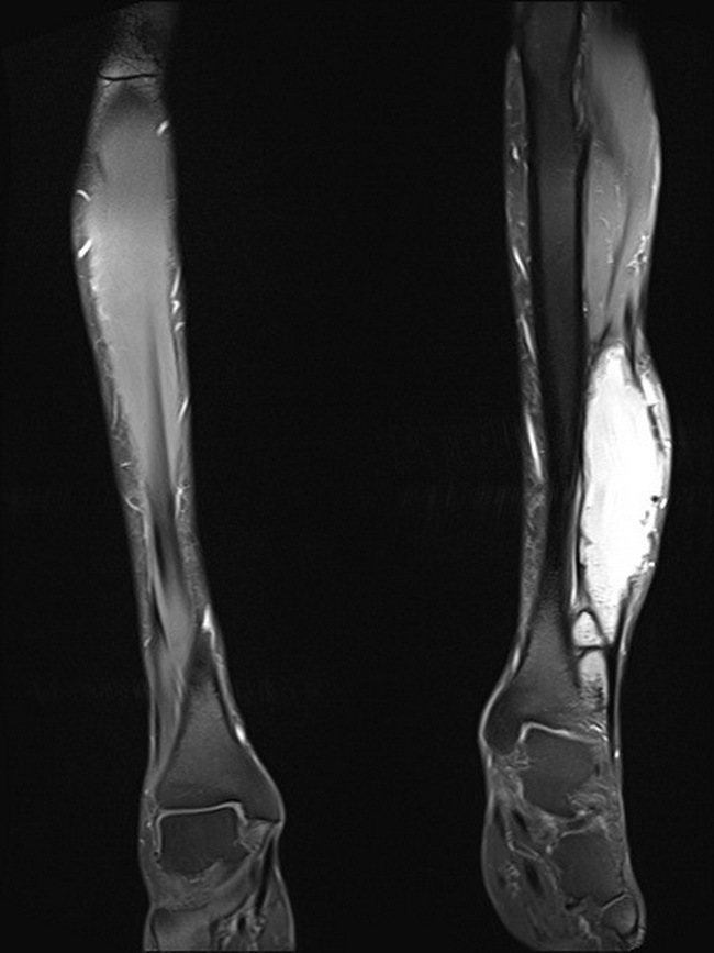
Myositis Ossificans - Imaging


Comments:
On MRI with contrast, myositis ossificans appears bright with a heterogenous appearance on T2-weighted images. The MRI findings may be worrisome in the initial stages as it appears ill-defined and infiltrative due to tissue edema. Case History (same as previous image): The patient was a 60 y/o male who presented with painful lower limb with multiple soft tissue masses. There was history of trauma resulting in fracture of tibia and fibula in the distant past. The lower limb shows multiple large sharply delineated lesions with mainly peripheral calcifications, consistent with late phase myositis ossificans. There is no edema or reaction in the surrounding tissues. Case courtesy of Dr Stan Buckens, Radiopaedia.org. From the case rID: 28650