
Long-segment Barrett Esophagus

slide 6 of 36


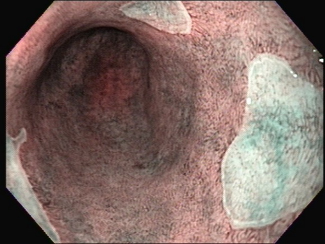
Comments:
Long-segment Barrett esophagus by narrow band imaging (NBI) endoscopy. Most of the distal esophagus is lined by metaplastic columnar epithelium (brown areas). The residual foci lined by squamous epithelium appear green. See image 4 for a discussion on NBI endoscopy. Image courtesy of: Pramod Malik, MD; used with permission.
slide 6 of 36