
Myeloma Kidney : Pathogenesis


Comments:
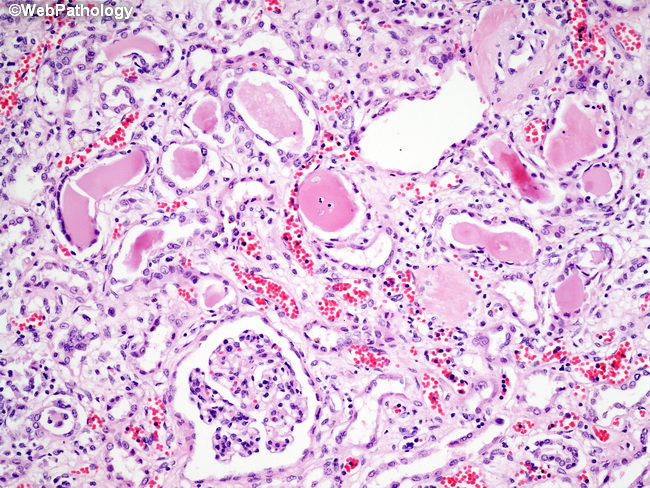
Myeloma patients develop tubulo-interstitial lesions due to light chain deposition in their kidneys, resulting in chronic renal insufficiency over time. Pathogenesis: Free light chains are filtered by the glomeruli but reabsorbed by the proximal tubular epithelial cells. In myeloma patients, the proximal tubules are unable to cope with the incoming load of excess light chains in the glomerular filterate. The unabsorbed light chains reach distal nephron where they form complexes with urinary glycoproteins and precipitate as large tubular casts (as shown here), blocking the lumen, and inciting an inflammatory response. In addition, light chains are directly toxic to the tubular epithelium. In up to 25% of myeloma patients, the renal damage is mediated by the development of amyloidosis of AL type due to deposition of free light chains (lambda type) in the interstitium. In some patients, there is deposition of kappa light chains in the glomerular basement membranes, mesangial matrix, as well as in the tubular basement membrane. Besides deposition of light chains, additional renal damage is caused by hypercalemia and hyperuricemia often encountered in myeloma patients.