
Hepatocellular Carcinoma in Hemochromatosis
Home
 Gastrointestinal
Liver
Liver Tumors & Tumor-like Lesions - II
Hepatocellular Carcinoma in Hemochromatosis
Gastrointestinal
Liver
Liver Tumors & Tumor-like Lesions - II
Hepatocellular Carcinoma in Hemochromatosis

slide 51 of 65


Comments:
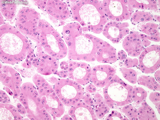
Higher magnification of the previous image shows predominantly acinar pattern with well-formed pseudoglandular spaces. The acini are formed by dilatation of bile canalicular-like structures elaborated by the tumor and often contain bile or proteinaceous material. The acinar pattern should not be mistaken for cholangiocarcinoma, which can be differentiated from HCC by immunoreactivity to MOC31, CK7, and CK19.
slide 51 of 65