
Meningioma - Radiographic Features

slide 5 of 60


Comments:
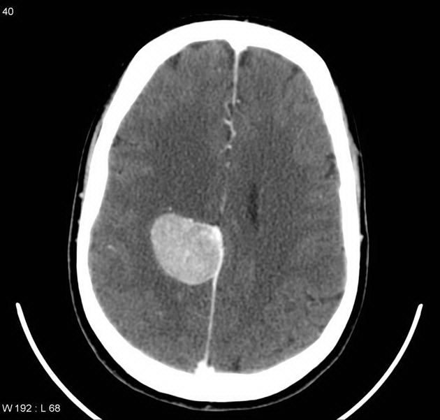
55 y/o male with meningioma arising in falx cerebri. The patient presented with left hemiparesis. On CT, most meningiomas appear hyperdense compared to the normal brain parenchyma. With contrast, most cases enhance brightly and homogenously as shown in this CT image. About 20% to 30% show calcification. Case reproduced with permission, courtesy of Dr. Frank Gaillard; Radiopaedia; Complete case is here.
slide 5 of 60