
Myositis Ossificans - Imaging


Comments:
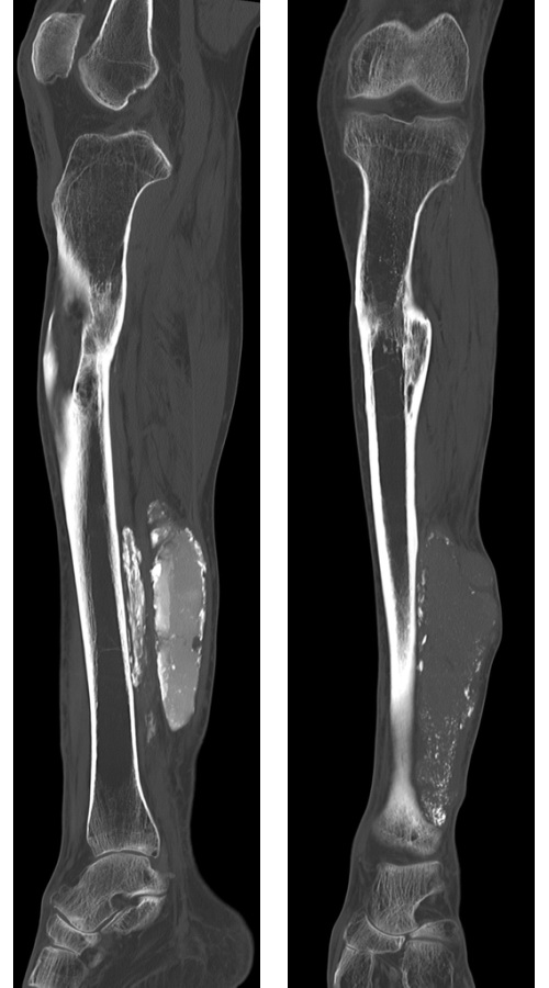
With CT, myositis ossificans appears well-circumscribed with peripheral ossification. In mature lesions, CT is the best imaging modality for delineating the zonal pattern of calcification. Case History: The patient was a 60 y/o male who presented with painful lower limb with multiple soft tissue masses. There was history of trauma resulting in fracture of tibia and fibula in the distant past. Note the soft tissue mass in the lower leg with peripheral coarse calcifications and radiolucent center in these CT images. The healed fracture in proximal tibia is also more easily appreciated. Fibula is not visualized. The clinical presentation, imaging studies, and morphology were diagnostic of myositis ossificans. Case courtesy of Dr Stan Buckens, Radiopaedia.org. From the case rID: 28650