
Myxopapillary Ependymoma : MRI Scan

slide 41 of 48


Comments:
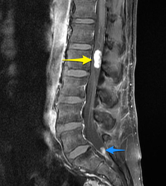
Myxopapillary ependymoma of the spinal cord. This 55 y/o male patient presented with neurogenic claudication. On MRI scan (T1 weighted, post-contrast), two tumors can be seen. The larger of the two (yellow arrow) is located at L2 level just below the conus medullaris. The smaller tumor (blue arrow) is located in the thecal cul-de-sac. The patient underwent laminectomy and resection of the larger tumor, both for symptom relief and diagnosis. Histology revealed typical myxopapillary ependymoma (WHO Grade 1). Case produced with permission, courtesy of Dr. Frank Gaillard. Radiopaedia. Complete case is here.
slide 41 of 48