
Hydatid Cyst : Liver

slide 4 of 32


Comments:
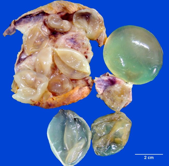
The patient was a 51 y/o female who presented with abdominal pain, nausea and fever. On examination, there was tenderness in the right upper quadrant. Liver function tests were within reference ranges; she had mild eosinophilia. Abdominal CT revealed a 9.7 cm hypodense cystic lesion in the right lobe of liver. Multiple well-defined round to oval cysts of various sizes were seen within the large cyst. The findings were diagnostic of Hydatid Cyst. The image shows parts of thick cyst wall and multiple daughter cysts, most of them collapsed and a few intact, ranging in size from 2 cm to 5 cm. Contributed by : Dr. Sanjay D. Deshmukh, Department of Pathology, Smt. Kashibai Navale Medical College, Pune, India.
slide 4 of 32