
Thymoma : Myasthenia Gravis


Comments:
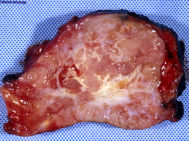
Thymoma and Myasthenia Gravis:About 30% to 45% of patients with thymoma develop myasthenia gravis. Conversely, myasthenia gravis patients show thymoma in 10% of cases, thymic hyperplasia in 65% of cases, and no obvious thymic pathology in 25% of cases. There are no obvious morphologic differences between thymomas that are associated with myasthenia and those that are not. Besides myasthenia gravis, thymomas are associated with a large number of immune-mediated systemic illnesses, including hypogammaglobulinemia, Graves disease, pure red cell aplasia, white blood cell aplasia, pernicious anemia, dermatomyositis-polymyositis, myeloma, autoimmune enteropathy, T-cell chronic lymphocytic leukemia and T-cell lymphoblastic lymphoma. The image shows a gross specimen of a thymoma. The tumor is pink-tan, solid, and composed of lobules separated by fibrous septa. About 80% of cases of thymoma are well-encapsulated. This case was minimally invasive - microscopic foci of tumor cells were seen infiltrating through the capsule into the surrounding mediastinal adipose tissue.