
Ovarian Fibroma : Gross Pathology


Comments:
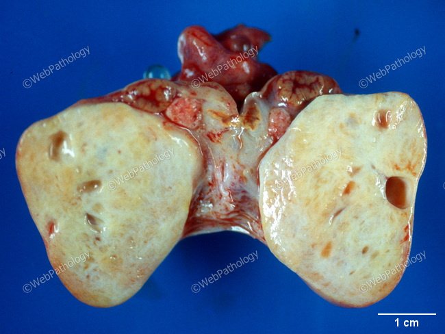
Gross Pathology: Ovarian fibromas are unilateral in 90% of cases. The average size is 6 cm (range - microscopic to >20 cm). Up to 20% may be pedunculated. They are well-circumscribed with a smooth lobulated outer surface and a homogenous, solid, firm tan-white cut surface. Some cases have a whorled appearance. The gross appearance is similar to leiomyoma, thecoma, Brenner tumor, and Krukenberg tumor. There may be myxoid or edematous change with pseudocyst formation (25% of cases; seen in this image) and calcification (10% of cases). Necrosis and hemorrhage are uncommon but may be present if the tumor undergoes torsion. Cellular fibromas tend to be larger in size (avg. diameter 12 cm) with a softer, fleshier, yellow-white cut surface as compared to conventional fibromas. They may also be associated with extraovarian adhesions and peritoneal implants. Ovarian fibromas in Gorlin syndrome are often bilateral, multinodular, multifocal and heavily calcified.