
Fibrous Hamartoma of Infancy : Microscopic

slide 3 of 60


Comments:
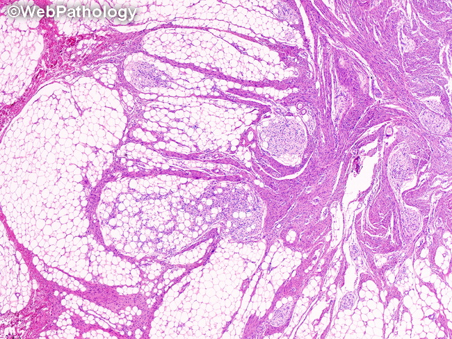
Microscopic Features: Fibrous hamartoma of infancy (FHI) consists of three distinct tissue components arranged in an organoid pattern - 1) haphazardly arranged, intersecting fascicles of cytologically bland fibroblastic/myofibroblastic cells; 2) mature adipose tissue; and 3) highly vascular nodules of primitive mesenchymal tissue. The relative proportion of the three components can vary widely from case to case, but the classic triphasic morphology is present at least focally. Mitotic activity is not increased. There is no necrosis. Two cases of FHI with sarcomatous areas have been reported. They showed high cellularity, brisk mitotic activity and high nuclear grade.
slide 3 of 60