
Dermatofibrosarcoma protuberans

slide 3 of 18


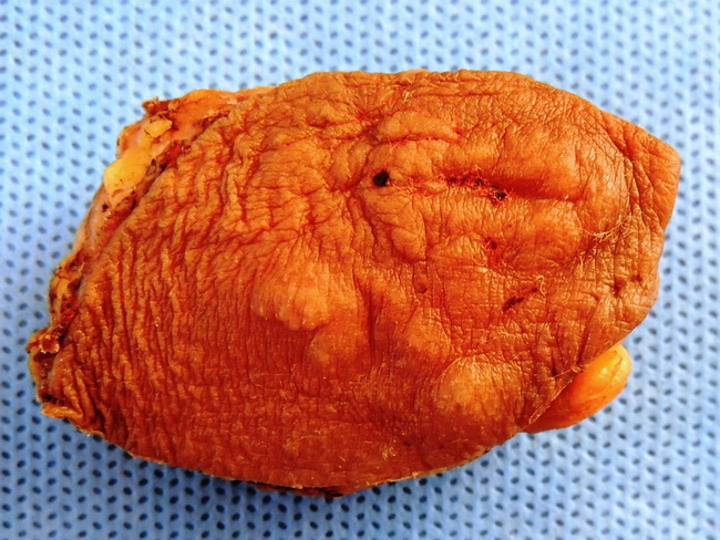
Comments:
This dermatofibrosarcoma protuberans presented in a 38 y/o female as a slowly-growing, non-tender, firm subcutaneous swelling above the lateral aspect of the right clavicle. The excised specimen revealed a fusiform swelling with scattered nodules on the overlying skin. The cut surface shows yellow-tan homogenous nodule involving the subcutis. The lesion appears to extend to the lateral surgical margins. Microscopic examination confirmed dermatofibrosarcoma. Case courtesy of: Dr. Sanjay D. Deshmukh, Professor of Pathology, Dr. Vithalrao Vikhe Patil Medical College & Hospitals, Ahmednagar, India.
slide 3 of 18