
Parathyroid Adenoma

slide 2 of 22


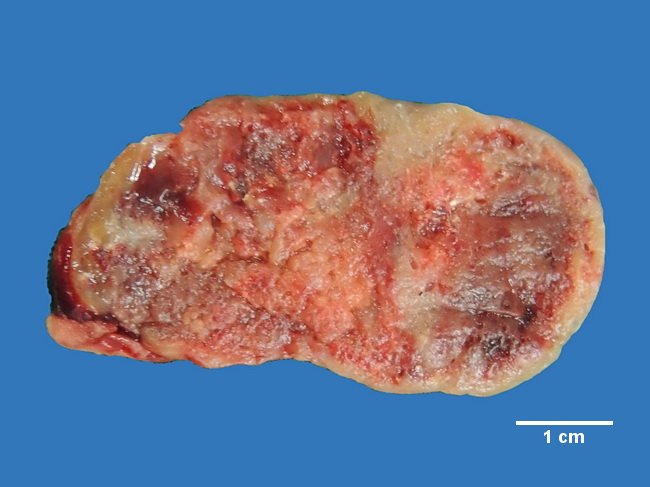
Comments:
The cut surface has greyish brown appearance with areas of hemorrhage. Sections showed a highly cellular vascular encapsulated tumor composed of chief cells, water-clear cells, and oxyphilic cells. Pleomorphic nuclei with degenerative type atypia were present. At the periphery, there was normal parathyroid tissue admixed with subcapsular adipose tissue. The clinical presentation in this case was primary hyperparathyroidism. The causes of primary hyperparathyroidism are parathyroid adenoma (85% to 90% of cases), primary hyperplasia (about 10% of cases) and parathyroid carcinoma (less than 1% of cases). Contributed by : Dr. Sanjay D. Deshmukh, Department of Pathology, Smt. Kashibai Navale Medical College, Pune, India.
slide 2 of 22