
Squamous Cell Carcinoma : Gross


Comments:
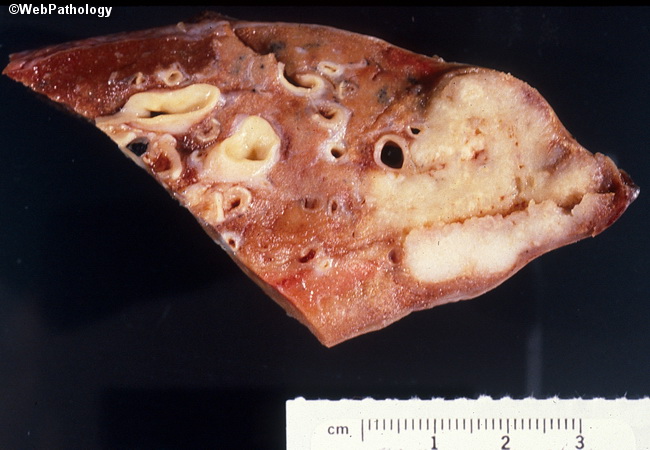
Squamous cell carcinomas usually arise centrally in a main or lobar bronchus and appear as a large hilar or perihilar mass. One third of cases arise peripherally or even subpleurally (as seen in this wedge resection specimen). The tumor surface is whitish-gray often speckled with black deposits of anthracotic pigment. There are soft, friable areas of necrosis which frequently cause central cavitation within the tumor. The tumors may form an endobronchial polypoid mass which may infiltrate through the bronchial wall into the lung parenchyma. The bronchial mucosa adjacent to the tumor frequently contains full spectrum of histologic changes, including squamous metaplasia, dysplasia, and carcinoma-in-situ. Obstruction of the bronchial lumen can result in atelectasis, obstructive bronchopneumonia, and bronchiectasis.