
Herpes Esophagitis


Comments:
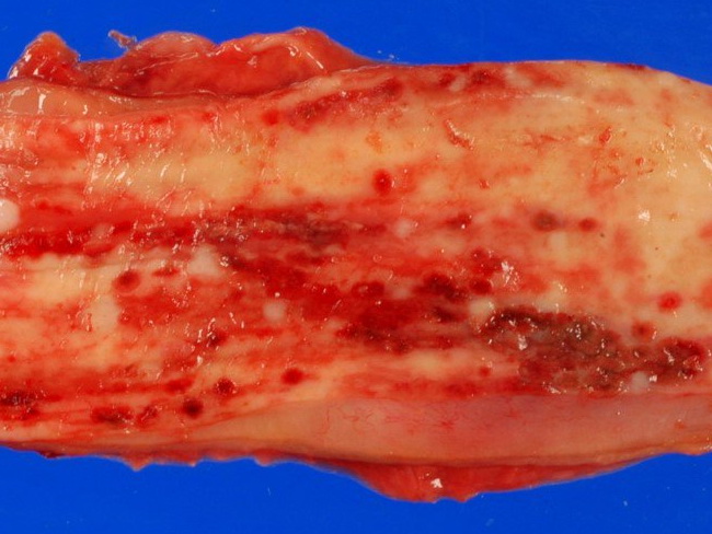
On endoscopy, herpes esophagitis shows discrete, shallow ulcers of variable sizes throughout the esophagus. The intervening mucosa may be normal or, more often, appears erythematous and friable. In severe cases, the ulcers coalesce and present as acute necrotizing esophagitis (aka "black esophagus"). Black Esophagus: Acute necrotizing esophagitis is a rare disorder that may be seen in a variety of clinical settings, including solid and hematologic malignancies, diabetic ketoacidosis, alcoholic intoxication, sepsis (including HSV and CMV), severe malnutrition, renal insufficiency, gastric volvulus, cardiovascular collapse, and vasculopathy etc. Endoscopy shows diffuse, circumferential black discoloration of the distal esophageal mucosa that stops abruptly at the gastroesophageal junction. This specimen of herpes esophagitis is from an autopsy case. Courtesy of: @PatholWalker; used with permission.