
Hemangioblastoma

slide 18 of 32


Comments:
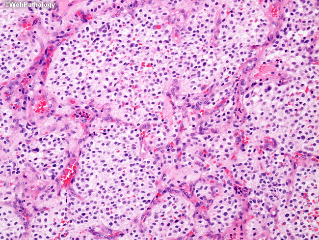
A subset of hemangioblastomas shows lobular clusters of tumor cells with less prominent cytoplasmic vacuolization. Such cases more closely resemble renal cell carcinoma. Immunohistochemistry can be used to work-up such cases. The stromal cells in hemangioblastomas are generally positive for inhibin and negative for cytokeratins, EMA, CD10, and pax-8. Renal cell carcinoma generally have the reverse profile.
slide 18 of 32