
Pineoblastoma

slide 15 of 20


Comments:
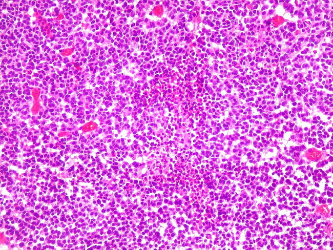
Pineoblastomas are composed of sheets of primitive-appearing cells with large hyperchromatic nuclei and minimal cytoplasm. Foci of necrosis can be seen in the center of the image. Courtesy of: Dr. Luciano de Souza Queiroz, Dept. of Pathology, Faculty of Medical Sciences, State University of Campinas (UNICAMP), Campinas, S�o Paulo State, BRAZIL. Additional images are here.
slide 15 of 20